If you are in the United Kingdom and have non-muscle invasive bladder cancer (NMIBC), previously known as superficial bladder cancer, you may be eligible to take part in our paid telephone/online interview. If you have a friend/relative who has NMIBC, please share this advertisement with them, as they could be eligible to take part in our paid telephone/online interview.
We are trying to find out about the aspects of treatment that matter to patients with NMIBC. Can you help? You can make your voice heard about your treatment priorities by taking part. Eligible participants will be paid £80 for completing the interview and £30 for providing proof of their diagnosis. The interview, including a brief online survey, will last up to 60 minutes.
If you are interested in participating or would like to receive further information about the study, please feel free to contact Nadine on nadine.koekemoer@evolve-fieldwork.com.
Alternatively you can leave your contact detail via the following link and we shall get in to contact with you: https://forms.gle/ZZvp1MM6p83fCy6R7
There is no evidence that the Bacille Calmette-Guérin vaccine (BCG) protects people against infection with COVID-19 virus. Two clinical trials addressing this question are underway, and WHO will evaluate the evidence when it is available. In the absence of evidence, WHO does not recommend BCG vaccination for the prevention of COVID-19. WHO continues to recommend neonatal BCG vaccination in countries or settings with a high incidence of tuberculosis.1
There is experimental evidence from both animal and human studies that the BCG vaccine has non-specific effects on the immune system. These effects have not been well characterized and their clinical relevance is unknown.2,3
On 11 April 2020, WHO updated its ongoing evidence review of the major scientific databases and clinical trial repositories, using English, French and Chinese search terms for COVID-19, coronavirus, SARS-CoV-2 and BCG.
The review yielded three preprints (manuscripts posted online before peer-review), in which the authors compared the incidence of COVID-19 cases in countries where the BCG vaccine is used with countries where it is not used and observed that countries that routinely used the vaccine in neonates had less reported cases of COVID-19 to date. Such ecological studies are prone to significant bias from many confounders, including differences in national demographics and disease burden, testing rates for COVID-19 virus infections, and the stage of the pandemic in each country.
The review also yielded two registered protocols for clinical trials, both of which aim to study the effects of BCG vaccination given to health care workers directly involved in the care of patients with COVID-19.4,5
BCG vaccination prevents severe forms of tuberculosis in children and diversion of local supplies may result in neonates not being vaccinated, resulting in an increase of disease and deaths from tuberculosis.6-8 In the absence of evidence, WHO does not recommend BCG vaccination for the prevention of COVID-19. WHO continues to recommend neonatal BCG vaccination in countries or settings with a high incidence of tuberculosis.
Bioprinting human tissue using specialised 3D printers promises to transform medicine, with implications for organ transplants, cancer treatment and antibiotic development.
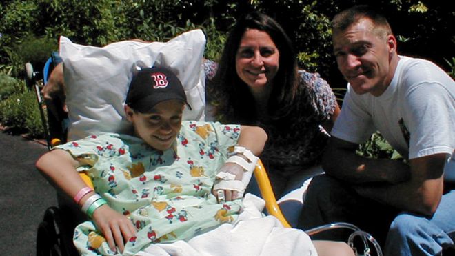
Luke Massella is one of about 10 people alive walking around with a replacement bladder that has been grown from his own cells.
He was born with a condition called spina bifida, which, from birth, left a gap in his spine.
By 10 years old, he had survived a dozen surgeries and beaten doctors’ initial expectations that he’d never walk. But then a malfunctioning bladder made his kidneys fail.
“I was kind of facing the possibility I might have to do dialysis [blood purification via machine] for the rest of my life,” he says. “I wouldn’t be able to play sports, and have the normal kid life with my brother.”
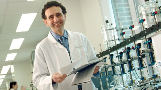
An enterprising surgeon, Anthony Atala at Boston Children’s Hospital, took a small piece of Luke’s bladder, and over two months grew a new one in the lab.
Then in a 14-hour surgical procedure he replaced the defective bladder with the new one.
“So it was pretty much like getting a bladder transplant, but from my own cells, so you don’t have to deal with rejection,” says Luke.
Rejection is when the body’s immune system attacks transplanted cells that come from another organism. Using tissue grown from a patient’s own cells helps combat this effect.
Luke went on to be a wrestling coach in the Connecticut public schools and now, at 27, runs events in the jewellery industry.
“Pretty much I was able to live a normal life after,” he says.
He underwent surgery 17 times before he was 13, but hasn’t had to since.
Dr Atala’s work involves bioprinting, using modified 3D inkjet machines to produce biological tissue.
His team has developed “eight cell-based tissues we put into patients,” he says, including engineered skin, urethras, and cartilage, all grown in the lab.
These engineered organs are going through clinical trials for approval by the US Food and Drug Administration.
“You need to know how to make these organs by hand, then the bioprinter is really a scale-up tool,” says Dr Atala, director of the Wake Forest Institute for Regenerative Medicine in North Carolina.
In other words, bioprinting would enable these organs to be made in an affordable, consistent, and precisely constructed way, he believes.
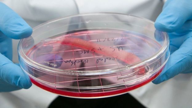
“Flat structures like skin” are easiest to print, he says. Then “tubular structures like blood vessels and urethras” are a little more complex, with “hollow non-tubular organs like bladders” harder still.
But hardest are “solid organs like hearts, lungs, and kidneys,” with “so many more cells per centimetre”.
For these highly complex organs bioprinters provide a precision that surpasses human hands, he says.
Pluripotent potential
Bioprinting has taken off following a dramatic discovery by Shinya Yamanaka and Sir John Gurdon, who a Nobel Prize for their work in 2012.
Adult ordinary cells can now be reprogrammed to make stem cells – called induced pluripotent stem cells – which can be used to make any other cell in the body.
“A lot has happened in the last couple of years,” says Steven Morris, chief executive of bioprinting start-up Biolife4d.
Mr Morris is working to bioprint a heart using these pluripotent cells over the next year. This will initially be a smaller version of the organ, he explains, but could eventually help pharmaceutical companies bypass testing trial drugs on animals, he says.
And ultimately, bioprinting organs from people’s own cells will solve the “huge lack of supply” in organs for transplant, says Mr Morris, and do away with the need for anti-rejection immunosuppressant drugs.
Specialist printers could even reproduce cancers tumours, giving doctors the chance to test “which treatment could specifically work on that patient,” says Erik Gatenholm, chief executive of Swedish start-up Cellink.
His firm has been given a €2.5m ($2.9m; £2.2m) grant from the European Union to develop these tumour-modelling printers.
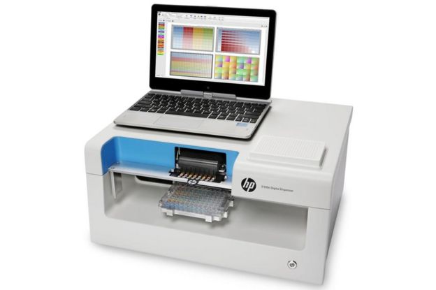
Bioprinters also give us a way of “quickly laying down small quantities of fluid to test if a new antibiotic would work for that specific patient,” says Annette Friskopp, vice president for specialty printing systems at the large tech firm HP in Palo Alto.
This could help tackle the growing and serious problem of antimicrobial resistance – the rise of “superbugs” traditional antibiotics can’t kill.
HP is partnering with the US Center for Disease Control to deploy printers in four regional labs in the US this autumn.
Inks and scaffolds
Printers of any kind need ink, and bioprinters are no different. “Bioink” is a gel that can be extruded through a printing nozzle and mimics the suspension lying between cells, called the extracellular matrix.
Both university labs and start-ups, such as Cellink, have been developing bioinks that can be used with many types of cells, says Ahu Arslan Yildiz, a biochemist who heads a research group at Izmir Institute of Technology in western Turkey.
And these “universal” bioinks are growing more and more “processable and easy to handle,” says Ms Yildiz, while also not being toxic.
Another breakthrough in the fast-developing field comes from Japan.
Most bioprinting uses a scaffold to hold cells in place. And once cells are “coaxed to a certain level, they begin to self organise and assemble,” says Arnold Kriegstein, director of the stem cells and regeneration medicine centre at the University of California, San Francisco.
They hope it will lead to more patients being cured while experiencing fewer side-effects.
Barry Dolling, 65, who has prostate cancer, is the first patient in the UK to be treated.
He told me: “I feel privileged and excited to be part of this. I volunteered to be part of the Prism trial and feel my treatment and the research will help others diagnosed with prostate cancer in the future.”
Pioneering
Prof Uwe Oelfke, who leads the joint MR Linac project at the ICR and Marsden, told me: “Ten years ago we did not think it would be possible to combine MRI and radiotherapy – this is a real step-change in technology.”
The machine has been specially designed so that the magnetic coils of the MRI and the X-ray beam of the linear accelerator do not interfere with each other.
It will be especially useful in treating tumours that shift in size and shape in the body, such as in the lung, bladder and bowel.
Dr Alison Tree, consultant oncologist at the Royal Marsden, described the new technology as “a dream come true” that would enable clinicians to deliver more targeted, higher doses of radiation, sparing healthy tissue.
“In lung cancer, we would like to give higher doses of radiotherapy but are limited because the tumour is often close to other vital structures in the chest,” she said.
“This allows us to see the cancer more clearly and make sure the radiation goes where it is needed and not where it can cause harm.”
Single treatment
The ability to give higher doses of radiation will enable patients to complete their treatment more quickly.
Mr Dolling will have 20 sessions of radiotherapy in the MR Linac. But it’s predicted that eventually it will be possible to cure some cancers in a single treatment.
The Royal Marsden will treat prostate, rectal, bowel, bladder, cervical and eventually lung cancer in the MR Linac.
The installation of the MR Linac was made possible by a £10m grant from the Medical Research Council, plus support from the Royal Marsden Cancer Charity.
A second machine, at the Christie in Manchester, will begin treating patients early next year.
Both hospitals are part of an international research consortium together with companies Elekta, which makes the MR Linac, and Phillips.
So far, the only other cancer centres to treat patients with the MR Linac are in the Netherlands, in Utrecht and Amsterdam.
The prospect of more accurately targeted radiotherapy, with fewer side-effects, will inevitably mean there will be high demand for access to treatment in the MR Linac.
Dr Tree said there would be several clinical trials and priority would be given to research that yielded the most benefit to patients.
There have been significant improvements in radiotherapy treatment in recent years – it already contributes to 40% of all the cases of cancer that are cured.
“Conversations: Let’s Talk About Bladder Cancer.” A 2014 instalment of this internet series features a discussion of the proper use of BCG (Bacille Calmette-Guerin). It features Dr. Donald Lamm and Dr. Michael O’Donnell, both bladder cancer experts.
New Bladder Cancer diagnostic test provides a unique combination of high sensitivity and negative predictive value
Lisbon, Portugal 21st October 2017. A new non-invasive urine test for bladder cancer providing a clear cut ‘yes/no’ result within three hours was launched at the 37th Société Internationale D’Urologie (SIU) meeting in Lisbon, Portugal, October 19-22, 2017. A study involving 577 patients, presented at an SIU symposium* today, showed the ADXBLADDER test was extremely reliable, demonstrating a sensitivity of 95% for higher risk cancers and a negative predictive value greater than 97%. The ADXBLADDER, produced by Arquer Diagnostics, received its CE Mark on 11th October 2017.
“ADXBLADDER is a real game changer in the field of bladder cancer testing. Our data demonstrates ADXBLADDER has one of the highest sensitivities and negative predictive values of any urine test for bladder cancer diagnosis and additionally offers innovative features, such as the result being unaffected by urinary tract infections (UTIs) and no requirement for samples to be sent away to specialist labs for analysis,” said Mr. Stuart McCracken, the study principal investigator.
“ADXBLADDER has the potential to make non-invasive testing for bladder cancer a clinical reality. It could help to diagnose patients earlier in the disease and offer dramatic improvements in quality of life and safety by reducing the need for cystoscopy and CT scanning,” said Mr. Tim Dudderidge, a study investigator.
The European Association of Urology currently recommends cystoscopy in all patients with haematuria and that cystoscopy cannot be replaced by cytology or any other non-invasive tests currently on the market. (1) The EAU guidelines also state that renal and bladder ultrasound may be used as part of the initial work-up and CT urography should be performed only in selected high-risk tumours. Nevertheless many CT urograms are performed in patients with visible and non-visible haematuria to exclude upper tract urothelial cancer, following guidance from the American Urological Association.(1) CT urograms, which use X-rays to construct detailed 2-D images of the bladder, have the disadvantage of exposing patients to ionizing radiation.
Cystoscopy, where the surgeon inserts a flexible cystoscope through the urethra to examine the inside of the urethra and bladder for signs of disease, is an undignified, invasive and uncomfortable procedure. Furthermore, cystoscopy exposes patients to the risk of urinary tract infections (UTIs), known to occur in up to 5% of cases undergoing cystoscopy (2), and used on its own misses up to 30% of bladder tumours. (3) (4) Cystoscopy also comes with additional health economic disadvantages including high costs, lengthy procedures and long waiting lists. For urine cytology, a sample of urine is examined under the microscope to check for cancer or precancerous cells. The downside of cytology is that a negative result does not exclude the presence of a tumour, cytology has a low sensitivity and interpretation is user dependent. (1)
“There is an urgent need for an alternative reliable investigation that provides reassurance but without the cost or risks of CT,” said Mr. Dudderidge, a Urology Surgeon from University Hospital, Southampton.
The ADXBLADDER test
The new ADXBLADDER test is based on measuring levels of a protein called MCM5, which is a marker of cells that are replicating, or still have the capability to replicate (i.e. are not terminally differentiated). Such cells are normally only found in basal layers of the epithelium, but when a mass of replicating cells is present (as occurs in bladder cancer), MCM5 cells are shed into body fluids. MCM5 in the urine is indicative of undifferentiated cells in an inappropriate location. Detection utilizes standard Enzyme Linked Immunosorbent assay (ELISA) methodology, using mouse monoclonal antibodies to identify MCM5 antigens.
An important feature of ADXBLADDER is that the test is not influenced by infections or inflammation. Bacteria do not contain MCM5 proteins and inflammatory cells are already differentiated and therefore do not express the MCM5 protein.
“It’s a simple test that provides a ‘yes/ no’ answer according to whether there’s been a colour change in the ELISA. An additional advantage is that the test uses standard ELISA kits, available in most hospital laboratories, and therefore won’t need samples sending away for analysis,” said Mr. McCracken, a Urology Surgeon from Newcastle University and Sunderland Royal Hospital, UK.
The ADXBLADDER study
In the ADXBLADDER blinded prospective study, between August 2016 and February 2017, 577 patients (318 male and 248 female) attending diagnostic haematuria clinics at six UK centres underwent ADXBLADDER ELISA testing. The results from the ADXBLADDER test were then compared to the current benchmark of combined results from cystoscopy, ultrasound and CT scanning.
The exclusion criteria for the study were patients with known kidney stones, or who had suffered previous bladder, prostate or kidney cancer.
The investigators found that 7.96% of the tests (n=46) were positive for cancer and 92.1 % (n=532) negative. The results showed:
The sensitivity for the combined high risk and muscle invasive groups was 95% (high risk groups 92%; muscle invasive groups 100%). [Sensitivity is the test’s ability to correctly detect patients who have bladder cancer].
The overall sensitivity for the study was 76%. (High risk 92%; muscle invasive group 100%; intermediate risk 75%; low risk 50%).
The overall specificity was 69%. [Specificity (also called the true negative rate) measures the proportion of negatives that are correctly identified as such (e.g. the percentage of haematuria patients without bladder cancer who are correctly identified by ADX as not having bladder cancer).
The negative predictive value (NPV) was 97%. [The NPV is the proportion of patients who test negative who are indeed true negatives].
“Having such a high negative predictive value is really important, because if your test is negative you can be 97% confident that the patient doesn’t have cancer,” said Mr. McCracken.
The investigators, he added, had not been unduly concerned by poorer performance in low grade disease. “The rate of progression of low grade bladder tumours is low and furthermore, these patients will re-present with symptoms in the interim period. What’s really important is that ADXBLADDER achieved very good sensitivity in high risk patients and those with muscle invasive disease, where cancer is likely to spread quickly, and prognosis is much poorer”, said Mr. McCracken.
Future uses of ADXBLADDER
At the symposium Mr. Dudderidge, considered some future uses for ADXBLADDER. ADXBLADDER, he said, could have an important role to play in diagnosing women with bladder cancer more quickly. Although men are known to be three to four times more likely to develop bladder cancer, women are more likely to die from the disease. (5) Studies show that 57% of men survive bladder for five years or more compared with just 46% of women. (6) (7) (8) Delayed diagnosis and women suffering from rarer forms of the disease are considered to be among the possible factors explaining this disparity.
One reason for the delay in diagnosis is that women are less likely to undergo assessment for haematuria due to suffering more commonly from urinary infections (which can cause similar bladder symptoms and blood in the urine just like bladder cancer). Studies suggest that around 50 to 60% of all women develop UTIs over the course of their life-time, and that UTIs occur more often in women than men at a ratio of 8:1. (9) (10) “The reality is that women who suffer from recurrent UTIs may not bother to go to the GP if they spot visible blood, and additionally GPs may just prescribe antibiotics and not investigate them further,” said Mr. Dudderidge.
The ADXBLADDER test, he suggested, could be positioned to differentiate bladder cancer from UTIs at an early stage and be offered to women who have unresolved blood in the urine after two weeks of UTI treatment. “The availability of an accurate, easy and affordable urine test will reduce the barriers to testing women with persisting urinary symptoms and permit earlier diagnosis of bladder cancer which could make a real difference to their outcomes,” said Mr. Dudderidge.
The test, he added, could additionally be used in patients who have been treated for high risk bladder cancer and now require regular monitoring for recurrence. “Such high risk patients can be required to undergo cystoscopy testing up to every three months initially and then for the rest of their lives. Given the choice most would adopt a urine test in a heart-beat if they thought it offered a safe alternative to invasive cystoscopy.”
Notes to editor
*The symposium ‘A Breakthrough in Non-invasive Bladder Cancer’ was held at 17.00 on Saturday 21st October at the Société Internationale D’Urologie meeting in Lisbon, Portugal. Interviews
Mr. Stuart McCracken and Mr. Tim Dudderidge are available for interview For further information contact:
Julia Kendrick
Kendrick PR
Mobile: +44 (0)7890711037
Email: julia@kendrickpr.uk
About Arquer Diagnostics Ltd ADXBLADDER, a non-invasive bladder cancer diagnostic test, is the first product to be launched by Arquer Diagnostics. Arquer Diagnostics, launched in 2015, is a company based on the development of non-invasive cancer diagnostic tests using the MCM5 (minichromosome maintenance protein) platform. The company arose from UroSens, a small enterprise founded by Nick Miller-Jones in 2005. The mission of Arquer Diagnostics is to deliver accurate, rapid, minimally invasive oncology diagnostic products. Arquer Diagnostics, who are headquartered in Sunderland, United Kingdom, are supported by an international medical advisory board. ADXBLADDER is currently commercialized in UK, France, Italy, Turkey and the Nordic region and will be further released to other European countries and worldwide regions in 2018.